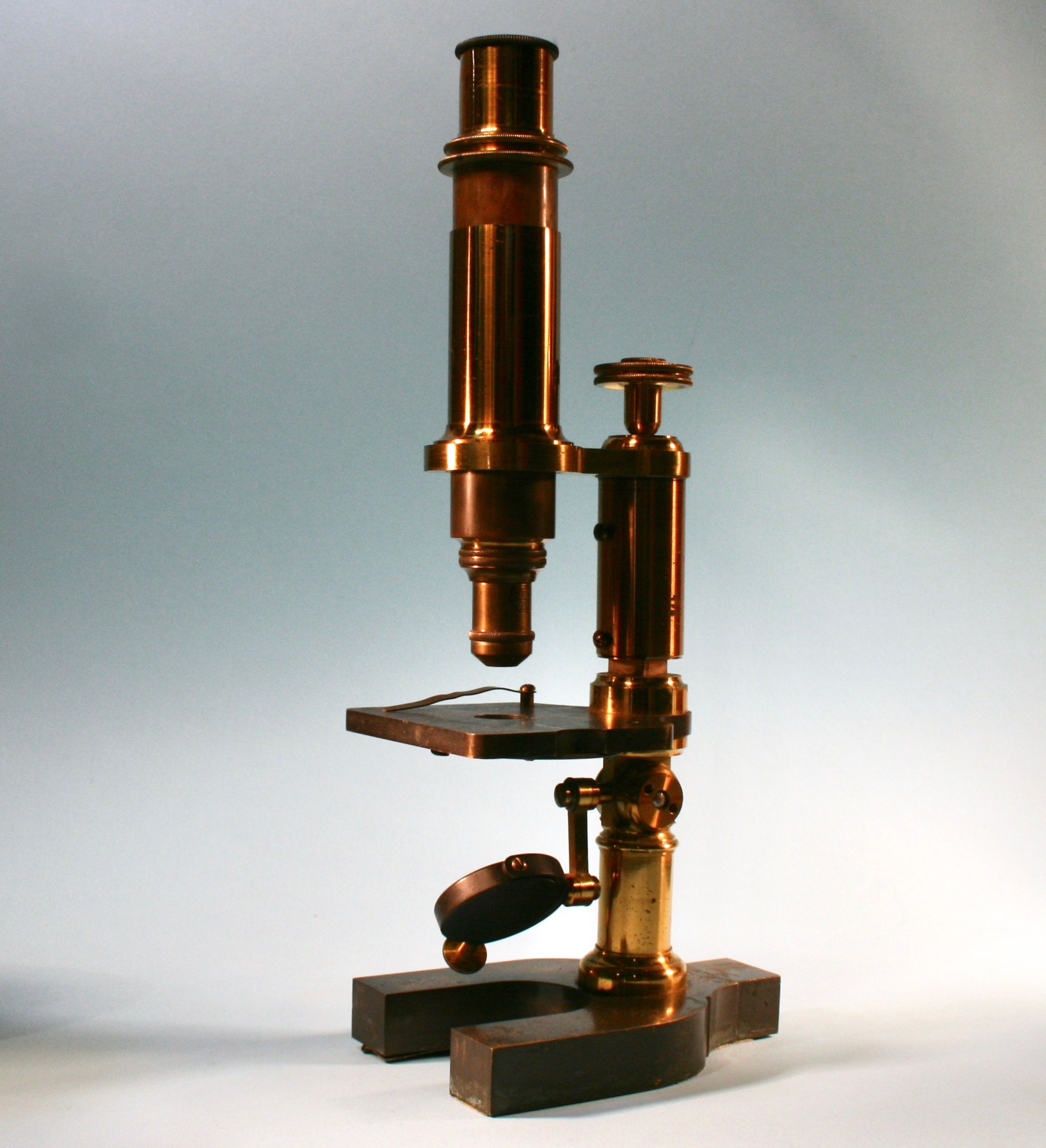

Title: Antique Microscope Scientific Lab Magnification Tool
Shipping: $29.00
Artist: N/A
Period: 19th Century
History: N/A
Origin: Central Europe > France
Condition: N/A
Item Date: N/A
Item ID: 66
This kind of articulated equipment Is Rare and unusual. Hand made Early 19th century. Lab Magnification Equipment. A Beautiful Antique Microscope. A microscope is an optical instrument used for viewing very small objects, such as mineral samples or animal or plant cells, typically magnified several hundred times. These hand made instruments were made two there exact custom specifications for each scientist In its day. A vintage microscope is a machinist work of art. There are many types of microscopes, and they may be grouped in different ways. One way is to describe the way the instruments interact with a sample to create images. The first kind of microscope to be invented was the optical microscope, which uses light to pass through a sample to produce an image. Although objects resembling lenses date back 4000 years and there are Greek accounts of the optical properties of water-filled spheres (5th century BC) followed by many centuries of writings on optics, the earliest known use of simple microscopes (magnifying glasses) dates back to the widespread use of lenses in eyeglasses in the 13th century.
Historians credit the invention of the compound microscope to the Dutch spectacle maker, Zacharias Janssen, around the year 1590. The compound microscope uses lenses and light to enlarge the image and is also called an optical or light microscope (versus an electron microscope). The simplest optical microscope is the magnifying glass and is good to about ten times (10x) magnification. The compound microscope has two systems of lenses for greater magnification, 1) the ocular, or eyepiece lens that one looks into and 2) the objective lens, or the lens closest to the object. Before purchasing or using a microscope, it is important to know the functions of each part.
Link: https://en.wikipedia.org/wiki/Microscope
A microscope is an instrument used to see objects that are too small to be seen by the naked eye. Microscopy is the science of investigating small objects and structures using such an instrument. Microscopic means invisible to the eye unless aided by a microscope. The first detailed account of the microscopic anatomy of organic tissue based on the use of a microscope did not appear until 1644, in Giambattista Odierna's L'occhio della mosca, or The Fly's Eye. The microscope was still largely a novelty until the 1660s and 1670s when naturalists in Italy, the Netherlands and England began using them to study biology. Italian scientist Marcello Malpighi, called the father of histology by some historians of biology. The performance of a light microscope depends on the quality and correct use of the condensor lens system to focus light on the specimen and the objective lens to capture the light from the specimen and form an image. Early instruments were limited until this principle was fully appreciated and developed from the late 19th to very early 20th century, and until electric lamps were available as light sources. In 1893 August Köhler developed a key principle of sample illumination, Köhler illumination, which is central to achieving the theoretical limits of resolution for the light microscope. This method of sample illumination produces even lighting and overcomes the limited contrast and resolution imposed by early techniques of sample illumination.