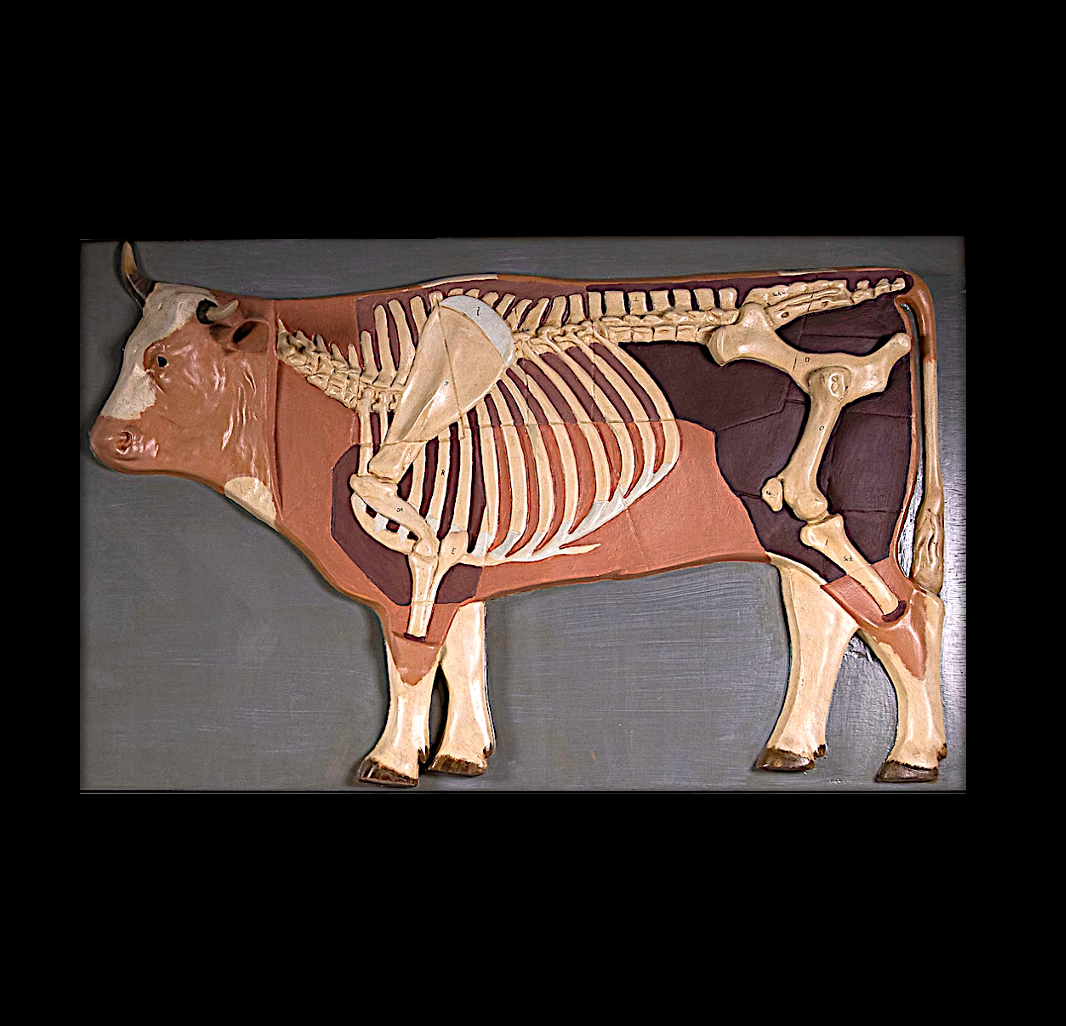

Title: Antique Classroom Early 20th Century Anatomical Model Cow Sculpture
Shipping: $29.00
Artist: N/A
Period: 20th Century
History: Art
Origin: North America > United States
Condition: N/A
Item Date: N/A
Item ID: 571
Consignment / call for availability and price / This is a captivating handmade internal diagram of a cow—an antique classroom early 20th-century anatomical model sculpture. These models, crafted for educational purposes in the 1920s and 1930s by Somso in Germany, are exceedingly rare. The wooden base measures 50-80cm (19.6-31.5 inches), and the overall height of the model is 3.50 inches. The materials used include wood and plaster. Originally published in the 1920s and 1930s, this model has undergone careful restoration by a restorer. While the surfaces of the flesh were once painted white, this was not the original color. Through the restoration process, the model has been returned to its authentic state, guided by reference photos. The colors surrounding the skin remain true to the original. Despite its age, the model is in good condition and retains its beauty, as evident in the accompanying picture. It served its purpose in education and stands as a testament to the craftsmanship of that era.
The history of anatomical models can be traced back to ancient times, with civilizations such as the Egyptians and Greeks creating rudimentary representations of the human body. However, the development of anatomical models as we know them today has a more structured and modern history. Renaissance Period (14th to 17th centuries): During the Renaissance, there was a renewed interest in human anatomy. Artists and anatomists collaborated to create detailed and accurate depictions of the human body. This era saw the emergence of wax anatomical models, often crafted by artists using the newly available medium of wax. These models were primarily used for teaching anatomy and surgery. 18th Century: Anatomical models continued to evolve during the 18th century. The use of wax models persisted, and there was an increasing emphasis on accuracy and realism. Anatomists and educators recognized the value of hands-on learning through the use of tangible models. 19th Century: The 19th century witnessed advancements in materials and production techniques. This era saw the introduction of papier-mâché as a medium for anatomical models, providing a more affordable alternative to wax. These models became popular in medical schools and universities for teaching purposes. 20th Century: The 20th century brought further innovation in anatomical modeling. Materials such as plastic and rubber became widely used, allowing for more durable and flexible models. Anatomical models also became more specialized, catering to specific medical disciplines. Advances in technology led to the development of virtual anatomical models, expanding the tools available for medical education. Contemporary Period: Today, anatomical models encompass a wide range of materials and technologies. Traditional materials like wood, plastic, and rubber are still used, but advancements in 3D printing and digital modeling have revolutionized the field. Virtual reality and augmented reality are increasingly being integrated into medical education, providing immersive and interactive learning experiences. Throughout history, anatomical models have played a crucial role in medical education, allowing students and professionals to study the intricacies of the human body in a tangible and informative manner. They continue to be indispensable tools in anatomy classrooms and medical training institutions worldwide.